Microsporidiosis
"last update:24 June 2024"
- Diagnosis
Clinical Signs
◾In C. gariepinus infected with Glugea sp. lesions characterized by whitish nodules (xenomas) embedded in kidney tissues containing viscous milky fluid (Abd Rabo, 2017). Some fish may carry parasites without demonstrating clinical signs, resulting in a subclinical infection. Nodules may be embedded in the connective tissue of the esophagus, intestinal submucosa, stomach, intestine and kidney (Fig. 3A, Abd Rabo 2017). In Saurida tumbil, Pleistophora aegyptiaca sp. nov. infection is associated with nodules (tumor like) in the peritoneal cavity, internal organs and within the skeletal muscle cells of fish produce grossly visible lesions (Fig. 3, Shehab El- Din, 2008). These nodules render the fish unmarketable (bdel-Ghaffar et al., 2012). Xenomas (cysts) may be embedded in all body organs, including muscles, liver, intestine, and stomach. Some marine fishes show white yellowish nodules caused by Glugea sp. and Pleistophora (Abdel-Mawla and Mohamed, 2010) whereas no nodules exist in Mugil cephalus.

Fig. 3: 3A) Xenomas embedded in the kidney as oval and small nodules. 3B) Saurida tumbil, showing Pleistophora aegyptiaca sp. nov. infection associated with nodules in the peritoneal cavity, internal organs and within the skeletal muscles.
▪️ Laboratory Diagnosis
Samples are taken by excising xenoma from affected organs which differ according to the pathogen species and the kind of infected fish.
▪️ Presumptive diagnosis
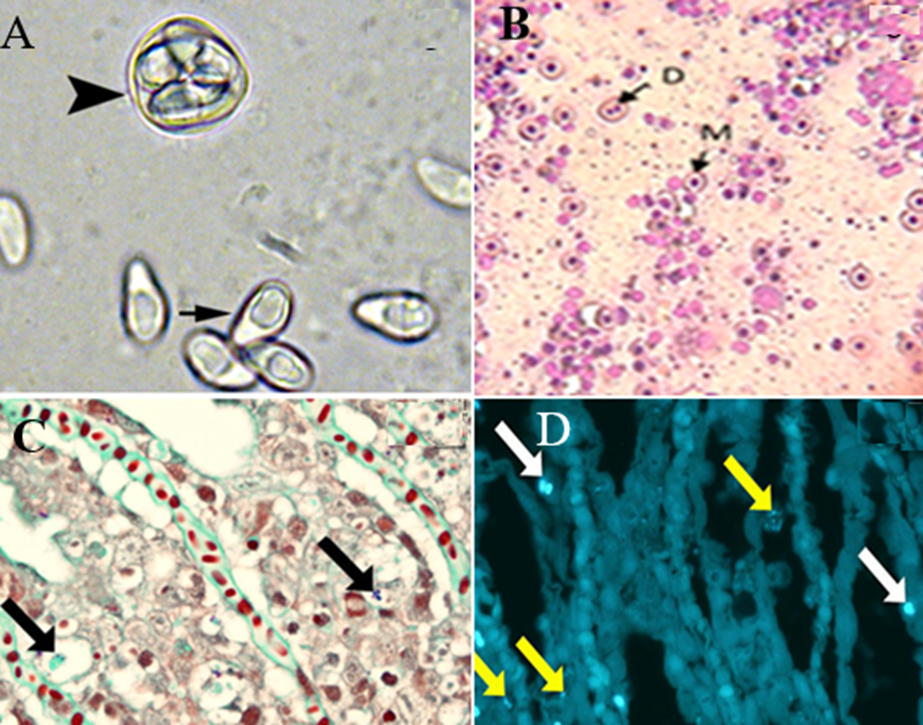
◾In fresh preparation from Yellow perch (Perca flavescens), spores appear oval to pyriform with an eccentrically located posterior vacuole and a sporophorous vesicle with several spores (arrowhead) of Heterosporis sutherlandae n. sp., (Fig. 4A) (Phelps et al., 2015). Wet mount from Glugea xenoma of C. gariepinus exhibits a typical egg-shaped spore of Glugea (Abd Rabo, 2017).
◾Staining xenoma contents with Geimsa stain revealed uninucleated meront (M) and dividing meront (D) (x40) (Fig. 4B) (Abd Rabo, 2017).
◾The microsporidian species can undergo spore discharge through the extrusion of the polar filament. This discharge can occur spontaneously (germination) or through incubation at various factors depending on the species. For instance, incubation at alkaline pH, acidic pH, or a pH shift from acid to alkaline or from alkaline to neutral. Dehydration through drying or hyperosmotic solutions followed by rehydration is another factor that can trigger spore discharge. Various cations including potassium, lithium, sodium, and cesium, and anions such as bromide, chloride, and iodide can also initiate spore discharge. Other factors that can trigger spore discharge include hydrogen peroxide, low dose ultraviolet radiation, and calcium ionophore A 23187 (Frixione et al., 1994. He et al., 1996).
▪️ Histological sections
◾Staining with conventional stains or Giemsa revealed that some spores appeared blue while others remained unstained.
◾Gram–Twort stain, both showing spores within the cytoplasm of degenerate epithelial cells (Fig. 4C) (Herreroet et al., 2020).
◾Staining with Calcofluor white fluorescent stain showing small (yellow arrows) and larger (white arrows) spores in the lamellar (Fig. 4D) (Herreroet al., 2020).

Fig. 4: 4A) spores appear oval to pyriform with an eccentrically located posterior vacuole (arrow) and a sporophorous vesicle with several spores (arrowhead) of Heterosporis sutherlandae n. sp. 4B) Staining xenoma contents with Geimsa revealed uninucleated meront (M) and dividing meront (D). 4C) Gram–Twort stain showing spores within the cytoplasm of degenerate epithelial cells. 4D) Calcofluor white fluorescent stain showing small (yellow arrows) and larger (white arrows) spores in the lamellar.
◾Transmission electron microscopy is a crucial research tool that is essential for a complete spore description.
▪️ Confirmatory diagnosis
◾PCR based assay; Samples are taken from the liver. The samples are collected from fish showing typical clinical signs. Liver samples are preserved in RNA later. DNA was extracted using the QIAamp DNA Mini Kit (Qiagen) following the manufacturer’s instructions. Fig. 5; The blasted sequence on the GenBank blast (NCBI) indicates that this sequence is related to fungus spp. (Alternaria Alternate) with similarity ~ 97%.

◾Molecular analysis of the rRNA genes including the ITS region of Glugea sp. uses species-specific designed primers.
◾Immunological methods Enzyme-linked immunosorbent assay (ELISA) and immunofluorescence assays can detect specific microsporidia antibodies in fish serum, indicating an infection.
◾Some species of microsporidia have been propagated in cell cultures.
