parasitic diseases of slaughtered food animals
| Site: | EHC | Egyptian Health Council |
| Course: | Food hygiene Guidelines |
| Book: | parasitic diseases of slaughtered food animals |
| Printed by: | Guest user |
| Date: | Tuesday, 24 December 2024, 4:35 AM |
Description
"last update: 13 Oct 2024"
- Acknowledgement
We would like to acknowledge the committee of National Egyptian Guidelines for Veterinary Medical Interventions, Egyptian Health Council for adapting this guideline.
Executive Chief of the Egyptian Health Council: Prof.Mohamed Mustafa Lotief.
Head of the Committee: Prof. Ahmed M Byomi
The Decision of the Committee: Prof. Mohamed Mohamedy Ghanem.
Scientific Group Members: Prof. Nabil Yassien, Prof. Ashraf Aldesoky Shamaa, Prof. Amany Abbass, Prof. Dalia Mansour, Dr Essam Sobhy
Editor: Prof. Nabil Yassien Prof. Heba Hussien Abdelnaem
- Abbreviations
NCC: Neurocysticercosis
C. bovis: Cysticercus bovis
C. cellulose: Cysticercus cellulosa
TB: Tuberculosis
F.H: Final host
IMH: Intermediate host
Kh: Conditionally approved after heat treatment
Kf: Conditionally approved after freezing treatment
T: Total condemnation
EMC: Encysted metacercaria
P/M: Postmortem
L.N: Lymph node
- Scope
|
III-Parasites not transmissible to man |
II-Parasites indirectly transmissible to man |
I- Parasites directly transmissible to man |
|
· Nematode (round worm): Ascaris and Lung worm · Cestodes (tape worm): Taenia ovis, Taenia hyaenha, Taenia hydatigena, and Taenia multicepes · Protozoa: Babesiaisis, Anaplasmosis, and Coccidiosis · Arthropode: Oestrus bovis (warble flies) and Oestrus ovis · Trematode: Fascioliasis |
Hydatid cyst |
Beef measle, Pork measle, Trichinella spiralis, Linguatula rhinaria, and Sarcosporidia |
|
- To categorize parasites which not transmissible to man - To identify the predilection seats of parasites which not transmissible to man - To identify the mode of infection of parasites which not transmissible to man - To differentiate between milk spots caused by immature ascaris or avian T.B in swine liver - To know the final and intermediate host of all parasites which not transmissible to man - To know the causative agent, postmortem lesion and judgement of butcher ̓s jelly - To differentiate between gide and false gide disease - To learn how to make judgements and control measures for all parasites which not transmissible to man - To learn forms of fasciola in different species. |
- To classify parasites which indirectly transmissible to man - To recognize the predilection seats of parasites which indirectly transmissible to man - To identify the mode of infection of parasites which indirectly transmissible to man - To know the final and intermediate host of parasites which indirectly transmissible to man - To learn how to make judgements and control measures for all parasites which indirectly transmissible to man
|
- To categorize parasites which directly transmissible to man - To study NCC (neurocysticercosis) - To differentiate between beef measle and pork measle - To differentiate between tongue worm and TB - To know the predilection seats for all parasites which directly transmissible to man - To identify the mode of infection of all parasites which directly transmissible to man - To recognize the final and intermediate host of all parasites which directly transmissible to man - To be able to make judgements and control measures for all parasites which directly transmissible to man - To learn how to test the viability of cysticerci - To study different forms of sarcosporidia (macroscopic and microscopic) - To study trichinoscopic examination of Trichinella spiralis |
- I- Parasites directly transmissible to man
|
|
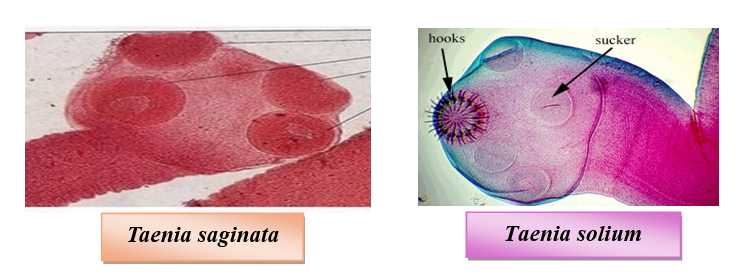
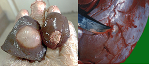
1. Beef measle |
2. Pork measle |
|
Cause |
Cysticercus bovis |
Cysticercus cellulosa |
|
F.H |
Man |
|
|
I.M.H |
Cattle and Buffalo |
Mainly in Pig and Man |
|
Judgement |
More than one cyst living or dead in an area of the size of a hand palm in different cuts of carcass is considered heavy infestation and requires T. Otherwise considered light and conditionally passed after freezing, boiling steaming, or pickling. |
Even one cyst living or dead detected in any part of the carcass or organ makes total condemnation (T).
|

3- Trichinosis (the smallest nematode)
Cause: Trichinella spiralis larvae
Host: Pig, wild boar, rat, mice, dog, man and cat. Ruminants, horses, and birds show natural immunity while camels can infest experimentally.
Resistance: Viable in decomposed meat for 2 year
Judgement: Even one cyst live or dead requires total condemnation
Control measures
- Pig flesh is trichinella-free
- Pork should be ground in a separate grinder
- Control rodents
- Proper cooking (at least 30 min at 100°C) of swill fed to pigs
Man
- Cooking: Meat color changes from pink to grey and easily separates muscle fibers.
- Freezing (-15 ° C for 20 days thickness less than 6 inches)
Irradiation: in countries allowing irradiation4- Sarcosporidia
• F.H: Ingestion of cysts (bradyzoites)
• I.M.H: Ingestion of feed with oocysts
• Predilection seats: Larynx- Esophagus- Diaphragm- Abdominal muscle- Lumber region muscle- Skeletal muscle (heavily infested cases)
• Judgement:
Localized affection condemnation of the affected part
Heavy infestation “T”
• Types (Forms) Sarcosporidia
|
Macroscopic |
Microscopic |
|
- Buffalo: Balbiana gigantea in the esophagus - Sheep: S. gigantea and S. medusiformis - Pig: S. porcifelis |
- Sheep: S. tenella - Cattle: S. blanchardi and cruzi - Pig: S. miescheriana - Man: • S. humnis (bovis humnis) and S. suihominis (F.H) • S. lindimanni (I.M.H) |


- II-Parasites indirectly transmissible to man
Hydatosis “Echinococosis”
Definition: Larval or cystic stage of Taenia echinococcus which is found in the small intestine of carnivorous
Cause: Hydatid cyst of Echinococcus granulosus “dog”- Hydatid cyst of Echinococcus mutilocularis “fox”
F.H: Dog and fox
I.M.H: Man, camel, cattle, sheep & pig in case of E. granulosus. Deer in case of E. multilocularis
Judgement:
Organ:
o Light infestation: Hygienic disposal of the cyst & part from surrounding tissue.
o Heavy infestation: Condemnation of organ


Carcass:
o Light infestation: Remove the affected part with hygienic disposal.The rest pass(no edema, emaciation, and muscular infestation
o Heavy infestation: Total condemnation (T)
Control measures: Carcass: Effective inspection and removal of affected part with hygienic disposal. Total condemnation of heavy-infested and emaciated carcasses with hygienic disposal. Man: Improve personal hygiene and Educational program.
- III. Parasites not transmissible to man
A. Nematode
1- Ascaris (toxocara)
Cause: Ascaris suum (suis) in pig. Ascaris ovis in sheep. Neoscaris(toxocara) vitulorm in calve.
P/M: Intestinal obstruction. Flesh: Characteristics odor (toxic substance and volatile acid)-poor. Liver: The early stage is congested and chronic cases make milk spots. Lung: Edema, hemorrhage, and parasitic pneumonia.
Judgement:
1. Condemnation of affected intestine and liver
2. Boiling and roasting test must be applied for detection of any abnormal odor & taste.

2- Lungworm Cause:
Dictyocaulus viviparous in cattle. Dictyocaulus filaria in sheep.
Metastrongylus elongatus in pig P/M:
Yellow or reddish brown foci (adult parasite)/surface of the lung just beneath
pleura. Catarrhal bronchitis and pneumonia (exudates contain the parasites).
Emaciated animal Judgement: Lung: o
Slight
infestation: condemnation of affected part o
Heavy
infestation: condemnation of lung Carcass:
If there is anemia and emaciation make T B- Cestodes 1- Sheep measle 2-Camel measle 3-Cysticerus tenuicollis 4-Coenurus cerebralis Cause C. Ovis C. cameli or
dromedarri C. tenuicollis (largest) Multiceps multiceps or Coenurus cerebralis Adult worm T. ovis T. hyaena T.hydatigena (marginata)
largest tape worm in dog T. multicepes (T. coenurus) Habitat Heart-
Masseter-Tongue- Diaphragm Heart- Masseter-Skeletal Ms- Liver Liver-Mesentery & its L. node- Omentum Brain &spinal cord F.H Dog Hyaena or dog Dog Dog I.M.H Sheep and goat Camel Ox, sheep, goat, pig, and camel Sheep, and goats. Rare in ox, horses, and man Judgement Organ: condemned Carcass: Light:
remove the affected part Heavy: (T) as c. bovis Organ: Light:
cyst. Heavy:
whole organ Carcass: A Early before emaciation remove head.
With emaciation (T) N.B: Gid=sturdy=turnsick= circling disease C- Arthropods 1- Oestrus bovis 2- Oestrus ovis Cause - Hypoderma bovis (cattle) - H. linatum - Sheep nasal fly / False gid Predilection
seat/ host - Muscle of belly (butcher's jelly) - S/C in goat - Carcass unmarketable - Sheep - Camels, dogs, and man (Sometimes) A/M and P/M - Swelling or
eroded skin back (Larvae protruding) - Cattle kick the
abdomen- erected tail - Paralysis
(lower body, legs) (spinal cord) - Hemorrhagic
edema -
Edema and
inflammation in s/c tissue around larvae (butcher ̓s jelly) - Symptom of brian irritation - Tozzing of head - Sneezing - Loss of appetite and emacaciation Judgement Trimming of the affected parts - Condemnation of
head - Carcass
approved (no emaciation) D- Trematode: Fascioliasis: Cause: Fasciola gigantica “Giant liver”, Fasciola hepatica, or Fascioloides
magna M.O.I: EMC on grass (animal, human). Not by adult fluke Acute Subacute Chronic - Immature - Haemorrhagic tracts - Swelling, congestion, Hg. on liver capsule - Soapy touch of muscle - Death from liver rupture - Differs from
the acute type in that symptoms are more protracted - Adult flukes in the bile ducts (mechanical
irritation) - Hard
liver and atrophy - The
cirrhotic areas (greyish color) Fasciola in different species: Cattle: Bile duct: Pipe
appearance/sand sound by knife. Liver tissue: Cirrhosis. Lung: Nodule (immature
fluke that failed to reach bile duct and liver) Sheep: Bile duct: Thickening &
dilatation. Liver tissue: Fibrosis. Lung: Rare nodule

Pig: Immature fluke encysted in
liver tissue & fail to reach bile duct. Judgement: Organ o Acute: Condemnation o Chronic: Light
infestation: Condemnation of the affected part. Heavy infestation: Condemnation
of the whole liver Carcass: o
Examined
for jaundice, emaciation & oedema "Cattle" o
Examined
for edema, emaciation, and jaundice "Sheep"
- References
· Kumar, S., Katoch, R., and Bhat, Z.F., (2014). Meat Borne Parasitic Zoonoses. In: Veterinary Parasitology. R. Katoch, R. Godara, A. Yadav, (Eds), Chapter 6, pp177-206. Satish Serial Publishing House, India.
· Bruschi, F., and Dupouy-Camet, J. (2022). Trichinellosis. In: Helminth infections and their impact on global public health. pp. 351-396. Cham: Springer International Publishing.
· Ortega Y.R., and Sterling, C.R. (2018). Foodborne Parasites. (Food Microbiology and Food Safety Series). 2nd ed., Springer.
· Xiao, L., Ryan, U., and Feng, Y. (2015). Biology of Foodborne Parasites; CRC Press: Boca Raton, FL, USA, Volume 1.
· Liu, D. (2018). Handbook of Foodborne Diseases. CRC Press, Boca Raton.
· Lawley, R., Curtis, L., and Davis, J. (2012). Parasites. In: The Food Safety Hazard Guidebook. Chapter 1.3, pp. 136-171. The Royal Society of Chemistry.
· Bruschi, F. (2021). Trichinella and trichinellosis. Elsevier.
· Campbell, W. (2012). Trichinella and trichinosis Springer Science & Business Media.